

- Accueil
- EN
- Studying at ULB
- Find your course
- fcsante
-
Share this page
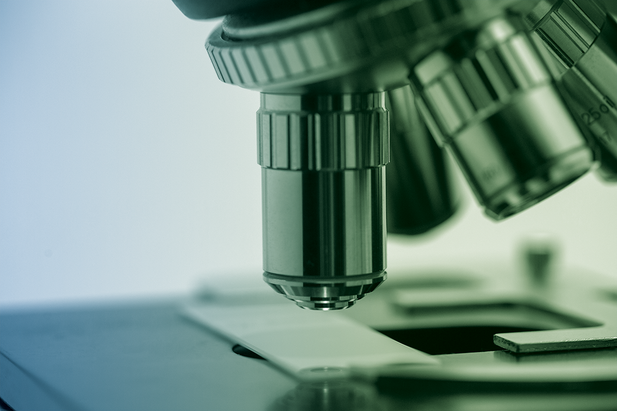
IMAGE-3.3 - introduction to fluorescence microscopy - sample preparation, microscopes, and image analysis
Short presentation
Acquire knowledge in processing and analysis of fluorescence microscopy imagesAccéder aux sections de la fiche
Call to actions
-
Programme titleIMAGE-3.3 - introduction to fluorescence microscopy - sample preparation, microscopes, and image analysis
-
Programme mnemonicFC-686
-
Programme organised by
- Centre de Formation Continue en Santé et Sciences de la vie
- Université libre de Bruxelles
-
Title typeformation continue
-
Open to returning studentsyes
-
Schedule typeDaytime
-
Languages of instructionenglish
-
Programme durationshort (2 to 5 days)
-
Category / TopicHealth - Biomedical and pharmaceutical sciences
-
Contact e-mail
-
Contact telephone
Presentation
Details
General information
Title typeformation continue
Programme durationshort (2 to 5 days)
Learning language(s)english
Schedule typeDaytime
Category(ies) - Topic(s)Health - Biomedical and pharmaceutical sciences
Organising faculty(s) and university(ies) Open to returning studentsyes
Tarifs- Job seeker rate: € 0.00
- ULB doctoral student rate (possibility of obtaining a grant): € 150.00
- Doctoral student rate other university: € 300.00
- Other public rate: € 500.00
- Industrial tarif: 700,00 €
Speakers
- Louise Conrard, PhD (ULB, CMMI)
- Egor Zindy, PhD (ULB, CMMI)
- Maud Martin, Pr (ULB, CMMI)
- Olivier Debeir, Pr (ULB)
Contacts
+32 71 37 86 96
Partner

Presentation
Programme objectives
- To obtain useful fluorescence images by optimizing sample preparation and understanding the microscope’s important elements and acquisition parameters
- To discover the various fluorescence microscopes and their specific domain of application
- To learn concepts and go-to methods for processing, analysing and quantifying images that were acquired with fluorescence microscopes
- To get familiar with ImageJ et CellProfiler, to learn how to use them, from basic usage to more advanced topics such as processing of image series and macro coding
Feel free to bring your own challenges in terms of samples or images to analyse
The course focuses on fluorescence microscopy images in life science, but the image processing concepts taught can be applied to any other scientific field.
Schedule
Daytime
- 2-day course- from 9:15 till 17:00
Calendar & registration
Target audience
- Biologists, researchers, technicians, and students wishing to use -or already using- fluorescence microscopy to answer biological questions, regardless to their specific field
- Staff working on bio-imaging platforms wishing to extend their knowledge.
- IT professionals, developers, image analysis specialists that are not yet familiar with software used in life sciences
Candidates will be selected based on their profile and the maximum number of seats available
Calendar & registration
Programme
PART 1: Learn the bases of fluorescence microscopy
- Which microscope should I use (to answer a specific biological question)?
- Sample preparation
- Microscopes operation and important image acquisition parameters
- Hands-on microscopes demonstration using Zeiss software or micro-manager
PART 2: Image processing and quantifications
- Concepts and methods of image processing:
- Bit depth, histogram adjustment, denoising and filtering, registration
- Concepts and methods of image analysis: Segmentation, tracking an object in image series, co-localisation of fluorescent objects
- Demo and exercises with ImageJ for routine image processing tasks, and CellProfiler for automated analyses and quantifications
- Overview of other useful software